HumanPhysiology.academy

home  

Lecture 14: Hemostasis: (Basic Principles of Coagulation)

Purpose: prevention of blood loss
(not to be confused with homeostasis)

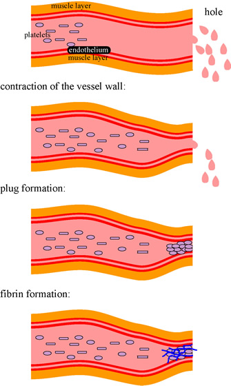
A. Sequence of events when a blood vessel is ruptured:


  1. spasm of the vascular wall
  2. formation of a platelet plug
  3. blood coagulation (i.e. coagulation is only a part of hemostasis)
  4. fibrolysis and growth of (fibrous) tissue (scar formation)


B. Vascular Spasm:

1.

Nervous reflex: due to pain and other impulses from the damaged area

2.

damage of the vascular wall also induces myogenic spasm

3.

Local humoral factors will also cause spasm: • from the traumatized tissue • from blood platelets (especially thromboxane A2)

4.

This vascular spasm may last from a few minutes to a few hours.

5.

There is a big difference between crushing and cutting wounds

6.

A spasm following crushing wounds last much longer and are much stronger. May even stop arterial bleeding.


C. Platelets:

These are 2-4 micron discs originating from the megakaryocytes in the bone marrow. They contain:

1.

no nuclei -> no reproduction

2.

actin & myosin -> are therefore contractile

3.

ER + Golgi apparatus -> can therefore synthesize enzymes

4.

Mitochondria -> makes ATP & ADP

5.

Platelets synthesize prostaglandins

6.

Platelets synthesize fibrin-stabilizing factor (for the next phase)

7.

Platelets synthesize growth factor (to repair damaged vascular walls)

8.

Platelets only attach to damaged cell membranes; not to normal endothelium


D. The Platelet Plug:

1.

Aggregation of swollen and sticky platelets

2.

Starts by platelets making contact with damaged cell (wall)

3.

The platelets become swollen and sticky

4.

Platelet secretion of ADP and thromboxane A2 activates other platelets (-> positive feed-back)

5.

In the initial stage, the plug is weak and loose, quite delicate, easily rubbed off (and that then induces new bleeding)


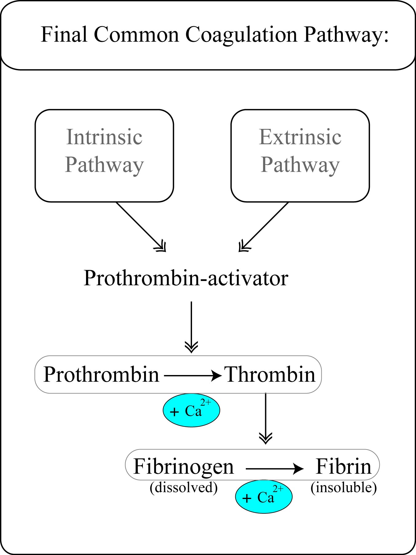
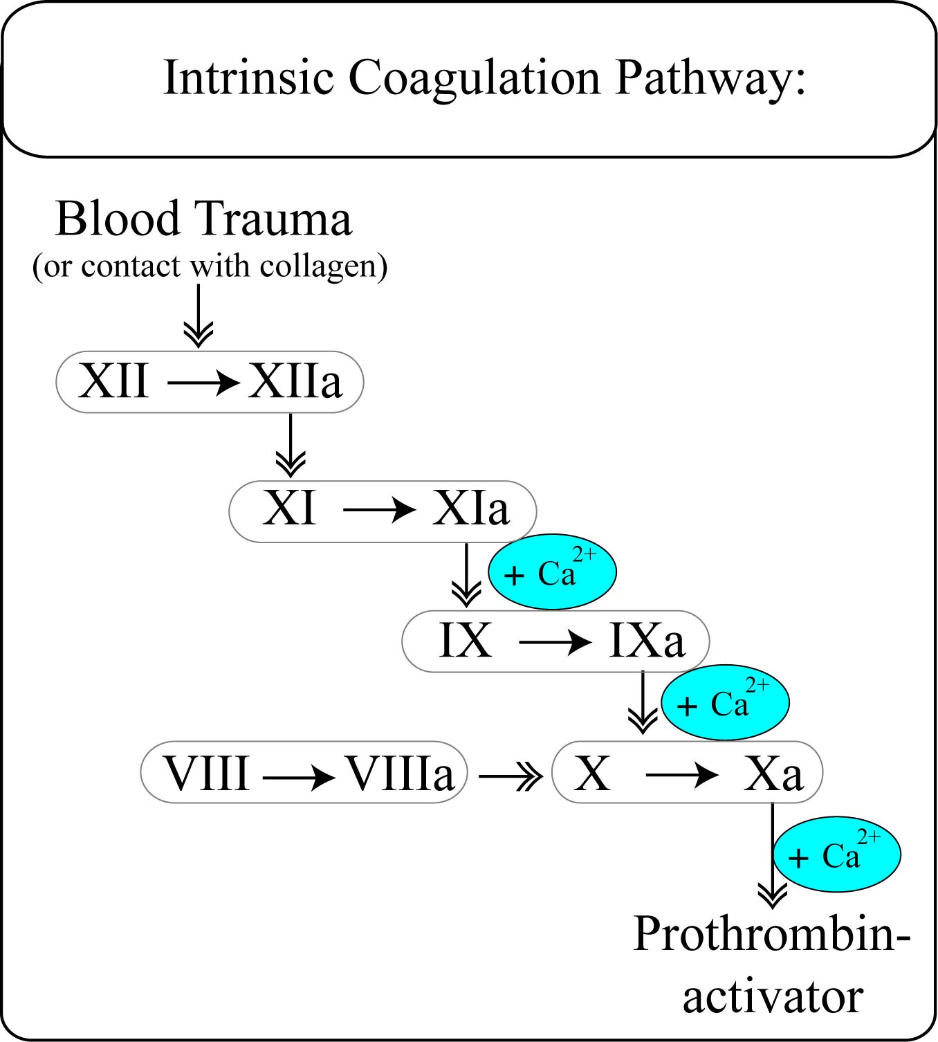
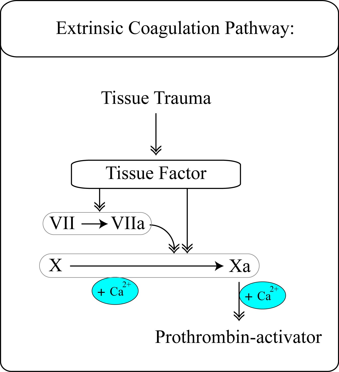
E. Blood clot formation: coagulation

1.

Development of fibrous threads into the platelet plug

2.

Takes 15 sec – 2 minutes

3.

Makes the plug strong and tight

4.

Also shows retraction, by pulling at the fibrin

5.

Uses fibrin stabilizing factor from the platelets

6.

Promoting coagulation -> procoagulants (such as blood factors)

7.

Inhibiting coagulation -> anticoagulants (such as heparine)

8.

In the circulation, there is a delicate balance between pro- and anti-coagulations. To much pro-coagulation and you induce intravascular clotting, to little and you induce bleeding.


F. The Blood Coagulation System:


The Blood Coagulation System consists of three branches:

1. an intrinsiek pathway (inside the blood)

2. an extrinsiek pathway (outside the blood; i.e. in the tissue)

3. a common pathway.


fff


The Clotting Factors and their Synonyms:

Clotting Factors:

Synonyms:

Fibrinogen

Factor I

Prothrombin

Factor II

Thromboplastin

Factor III (tissue factor)

Calcium

Factor IV

Factor V

Proaccelerin; labile factor

Factor VII

SPCA; stable factor

Factor VIII

antihemophilic factor A

Factor IX

antihemophilic factor B

Factor X

Stuart factor

Factor XI

plasma thromboplastin antecedent

Factor XII

Hageman factor

Factor XIII

Fibrin-stabilizing factor

Prekallikrein

Fletcher factor

Platelets


G. Lysis:

1.

After stopping the bleeding, the trauma must be repaired permanently. This is done mainly by plasmine that digests the fibrin threads.

2.

Plasmine is already in the clot, in the form of plasminogen. Plasminogen needs to be activated by TPA (tissue plasminogen activator) that is already available in injured tissue and injured endothelium.

3.

TPA and derivatives are now commercially available to treat clots (coronary clotting especially)


H. Excessive Bleeding: due to deficiency of clotting factors

1.

Vit. K deficiency: intestinal or liver diseases

2. Hemophilia:

a. shortage of factor VIII (85%)

b. shortage of factor IX (15%)

c. both are X-chromosome linked -> mother carrier; males affected

3.

Thrombocytopenia (platelet deficiency; thrombocytopenia purpura) -> makes the daily wear and tear evident in all our tissues.


I. Thrombo-embolic conditions:

1.

There is a big difference between a thrombus and an embolus.

2.

Thrombus formation is caused by:

a) roughened endothelial surface (arteriosclerosis / infection / trauma)

b) very slow moving blood (femoral thrombosis when immobilized in bed) -> classic story: first attempt at standing up after weeks of immobilization -> acute death

3.

Disseminated intravascular coagulation: in septic shock due to endotoxins.


Previous:
Home
Next:


© HumanPhysiology.academy 2014